Scapholunate ligament injury
A scapholunate ligament injury is a tear of the main ligament that joins two wrist bones, the scaphoid and the lunate. It most often happens after a fall on the hand and causes pain on the back of the wrist, sometimes a clunk and a loss of strength. Wrist arthroscopy (keyhole surgery) makes it possible both to confirm the injury and to treat it; if left untreated, a complete tear can progress to arthritis.
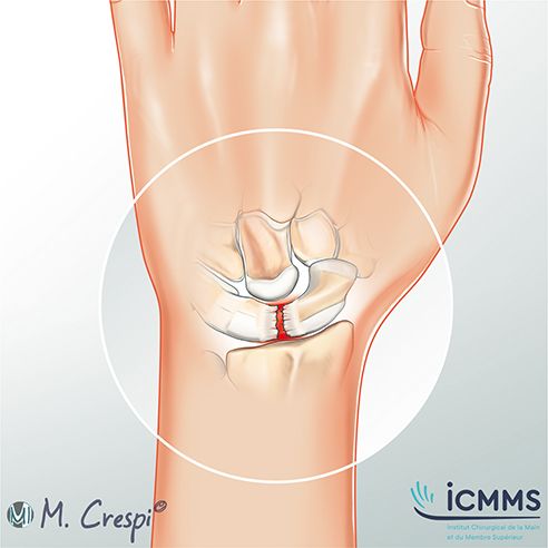
What is the scapholunate ligament?
The wrist is made up of two rows of small bones (the carpus) that move in a coordinated way. The scapholunate ligament connects the scaphoid, on the thumb side, to the lunate, in the centre of the wrist. It is one of the main stabilisers of the carpus: when it is torn, the scaphoid and the lunate move apart and tilt in opposite directions, which disrupts the mechanics of the wrist.
Injuries range from simple stretching to complete rupture, with or without damage to the neighbouring ligaments. The most serious form is the perilunate injury: during a violent injury, the ligaments give way all around the lunate, up to dislocation of the carpus, sometimes with associated fractures (of the scaphoid in particular). This is a surgical emergency, which is still too often missed.
What are the symptoms?
- Pain on the back of the wrist, on the thumb side, after a fall or a twisting injury
- Swelling in the days following the injury
- Loss of strength for gripping or carrying
- Pain when putting weight on the hand (press-ups, getting up)
- A painful click or clunk during certain movements
- A feeling that the wrist is unstable
How is it diagnosed?
- Clinical examination: the surgeon looks for pain at a precise point between the scaphoid and the lunate and performs specific manoeuvres, such as the Watson test, which provokes a painful clunk of the scaphoid
- X-rays of both wrists, including with the fist clenched: they can show an abnormal gap between the scaphoid and the lunate, or tilting of the bones, in significant tears
- CT arthrogram (CT scan with injection of contrast dye into the joint) or MRI scan: these show the ligament and any associated injuries
- Wrist arthroscopy: a small camera inserted into the joint makes it possible to see the ligament directly, assess the severity of the injury and treat it at the same time
The initial X-rays may be normal: a wrist that is still painful several weeks after a fall should be reassessed.
What are the non-surgical treatments?
Sprains and partial injuries without instability can be treated with:
- immobilisation in a splint for a few weeks
- painkillers and anti-inflammatory medication
- proprioceptive rehabilitation: exercises that strengthen the stabilising muscles of the wrist and improve control of movement
This treatment is often insufficient when the tear is complete.
When is surgery needed?
Surgery is offered in the case of a complete tear, instability, or pain that persists despite non-surgical treatment, and as an emergency in the case of a perilunate injury. Repairs are simpler in the first weeks after the injury: seeking advice early widens the options.
How is the operation performed?
The technique depends on how long ago the injury happened and how severe it is, as well as on the condition of the cartilage:
- Arthroscopy: it confirms the injury and allows tightening of the joint capsule (capsulodesis)
- Ligament reconstruction (ligamentoplasty): if the ligament is too damaged, it is replaced with a piece of tendon; this procedure can be assisted by arthroscopy
- Perilunate injury: reduction of the dislocation (putting the bones back in place), repair of the ligaments and fixation of the fractures, with temporary screws (an arthroscopically assisted technique described in a study co-authored by Dr Cievet-Bonfils: Hand Surg Rehabil 2026)
- Established arthritis: when long-standing instability has damaged the cartilage, a partial wrist fusion (arthrodesis, joining some of the carpal bones together) or other palliative procedures are discussed (see wrist arthritis)
The operation is done under regional anaesthesia of the arm, most often as a day case.
After the operation
- Immobilisation in a thermoplastic splint is worn for 6 weeks
- Any pins (K-wires) or screws are removed in the operating theatre once the ligaments have healed, at 6 weeks (under regional anaesthesia for the screws)
- Rehabilitation is gradual, aiming first at movement, then at strength and proprioception (the sense of joint position)
- Return to sport and heavy manual work usually takes several months
Some loss of movement is possible after a reconstruction; the aim is a stable, less painful wrist.
Frequently asked questions
My X-rays are normal: can I still have a scapholunate injury?
Yes. Partial injuries and some recent tears do not show on plain X-rays; a CT arthrogram, an MRI scan or an arthroscopy is then needed.
What happens if a complete tear is not treated?
The scaphoid and the lunate continue to work poorly together, which gradually wears the cartilage and leads over several years to wrist arthritis (known as SLAC: *scapholunate advanced collapse*).
Is wrist arthroscopy a major operation?
No. It is done through a few incisions of a few millimetres, as a day case; it is mainly the procedures performed during the arthroscopy that determine how long the wrist is immobilised.
What is a perilunate dislocation?
It is the most serious form of ligament injury of the carpus, in which the bones dislocate around the lunate after a violent injury; it requires reduction and surgical repair as an emergency.
Consultations in Lyon
Dr Maxime Cievet-Bonfils sees patients at:
- ICMMS – Centre Eiffel, 18-20 rue Tronchet, 69006 Lyon (3rd floor)
- Clinique de l'Infirmerie Protestante, 3 chemin du Penthod, 69300 Caluire-et-Cuire (Building D, 4th floor)
Secretary (direct line): +33 4 87 25 83 35 · Consultations in French or English.
This page provides general information. It does not replace a consultation: every situation is different.